Foot fusion surgery is a crucial procedure for those suffering from severe foot pain or deformities. This surgery aims to relieve discomfort and restore function by permanently joining two or more bones in the foot. Understanding the process and outcomes of this surgery can be daunting, but visual aids can provide valuable insights.
Pictures of foot fusion surgery offer a glimpse into the surgical techniques and the anatomy involved. They help demystify the procedure for patients and their families, showcasing what to expect during recovery and the potential benefits of the surgery. By examining these images, individuals can gain a clearer understanding of their treatment options and the transformative impact of foot fusion surgery on mobility and quality of life.
Overview of Foot Fusion Surgery
Foot fusion surgery, also known as arthrodesis, plays a crucial role in treating severe foot pain or deformities. This procedure involves the strategic joining of specific bones in the foot to reduce discomfort and enhance functionality. Surgeons select the affected joints for fusion based on individual conditions, utilizing various techniques to promote bone healing.
The primary objective of foot fusion surgery is to eliminate pain caused by conditions like arthritis, fractures, or structural deformities. By permanently uniting bones, the procedure restricts joint movement, which ultimately reduces pain. Patients often experience improved stability and function, facilitating a return to daily activities.
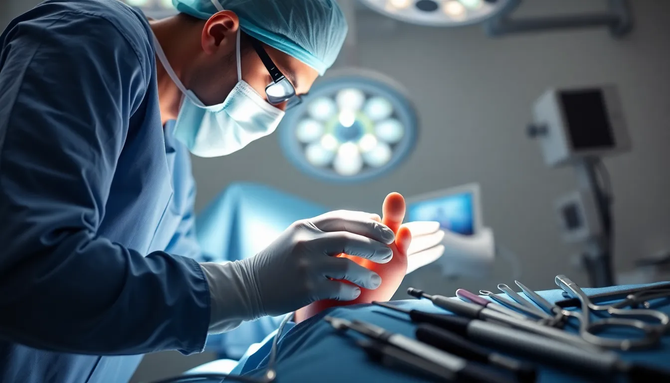
Pictures of the surgery provide essential insights into the surgical process, equipment utilized, and typical post-operative conditions. Such visual aids assist patients and their families in understanding the complexities of the procedure, from initial incision locations to the final steps of bone alignment. They offer a realistic perspective on recovery expectations and the various stages of healing.
Typically, recovery involves a gradual increase in activity, with initial immobilization through casts or braces. Rehabilitation plays a vital role in restoring strength and range of motion after surgery. Clear communication with healthcare providers assists patients in navigating their recovery journey effectively.
Importance of Visual Documentation
Visual documentation plays a crucial role in foot fusion surgery, enhancing the overall understanding of the procedure for patients and healthcare providers. Pictures provide clarity and context, making complex information more accessible.
Benefits of Pictures in Medical Education
Pictures serve as powerful educational tools in medical settings. They illustrate surgical techniques, anatomy, and potential complications effectively. Medical students and professionals benefit from visual aids, as they can reference real images that depict actual surgical scenarios. Research shows that visual learning increases retention rates and comprehension, essential for mastering surgical procedures like foot fusion.
Enhancing Patient Understanding
Pictures significantly improve patient comprehension of foot fusion surgery. Patients grasp the surgical process, visualize incisions, and better understand post-operative care. Visual documentation helps alleviate anxiety by setting realistic expectations for recovery and outcomes. Patients and their families can refer to images illustrating various stages, from preparation to rehabilitation, which supports informed decision-making. This transparency fosters trust between patients and healthcare providers, ultimately enhancing the surgical experience.
Types of Foot Fusion Surgeries
Foot fusion surgeries encompass various procedures tailored to address specific foot conditions. Understanding these techniques helps patients make informed decisions about their treatment options.
Common Procedures
- Lapidus Procedure: This technique fuses the first metatarsal and the medial cuneiform bone. It treats conditions like hallux valgus (bunions) and enhances stability in the big toe.
- Subtalar Fusion: This procedure fuses the subtalar joint, located between the talus and calcaneus bones. It’s effective for treating severe arthritis and instability in the hindfoot.
- Midfoot Fusion: Midfoot fusion connects bones in the midfoot region. It’s commonly performed for conditions like Charcot foot disease and arthritic joints.
- Ankle Fusion: This surgery unites the tibia and fibula with the talus bone at the ankle joint. It addresses severe ankle arthritis or joint damage, providing relief and function.
- First MTP Joint Fusion: This involves fusing the metatarsophalangeal joint of the big toe. It’s primarily used for advanced arthritis impacting toe movement.
Rare Surgical Techniques
- Triple Arthrodesis: This complex procedure fuses three joints in the hindfoot: the subtalar, calcaneocuboid, and talonavicular joints. It addresses severe deformities and arthritis.
- Talonavicular Fusion: This involves fusing the talonavicular joint, often for conditions resulting from advanced flatfoot deformities or instability.
- Cuneiform Fusion: This technique fuses the cuneiform bones to correct specific deformities or arthritis impacting the midfoot region.
- Distal Metatarsal Osteotomy with Fusion: This combines an osteotomy with fusion at the metatarsal heads, helping to reshape and stabilize the foot structure.
Each type of foot fusion surgery serves specific conditions, offering solutions based on individual patient needs.
Analyzing Pictures of Foot Fusion Surgery
Analyzing pictures of foot fusion surgery provides crucial insights into the procedure, recovery, and educational value for patients and providers. Visual aids enhance understanding of surgical techniques and outcomes.
What to Look For in Surgical Images
When examining surgical images, focus on several key elements:
- Incision Site: Observe the location and type of incision made during the procedure; this indicates the surgical approach taken.
- Bone Alignment: Assess the positioning of the bones involved in the fusion; proper alignment is critical for successful outcomes.
- Implants and Hardware: Identify any screws, plates, or other devices used; these support the fusion and stability of the joint.
- Post-Operative Condition: Look for signs of swelling or bruising; normal post-operative changes can inform about the expected recovery process.
- Healing Progress: Review images from various stages of recovery; these show the evolution of healing and how the foot may appear as it recovers.
Understanding Surgical Outcomes Through Pictures
Pictures play a vital role in comprehending surgical outcomes. They illustrate the transformation from pre-operative conditions to post-surgery results.
- Pain Relief: Display images that show improved alignment and joint function; this correlates with patient reports of decreased pain.
- Mobility Restoration: Document changes in foot function over time; improved mobility indicates successful surgery.
- Visual Comparisons: Utilize before-and-after images to showcase the effectiveness of different surgical techniques; this aids in decision-making for prospective patients.
- Rehabilitation Stages: Capture stages of rehabilitation to depict expected recovery timelines; these images help patients set goals for their recovery journeys.
- Patient Confidence: Showcase images of successful outcomes; positive results foster confidence in patients contemplating similar procedures.
Patient Experiences and Testimonials
Patient experiences provide valuable insights into foot fusion surgery and its impact on quality of life. Many patients express relief from chronic pain and improved mobility following the procedure. They describe returning to activities previously hindered by foot pain, such as walking, running, or playing sports.
Testimonials often highlight the importance of clear communication with healthcare providers before and after surgery. Patients appreciate when surgeons thoroughly explain the procedure, recovery timeline, and rehabilitation expectations. Knowing what to anticipate helps manage anxiety and promotes confidence throughout the recovery process.
Patients commonly report varying recovery experiences, emphasizing that initial immobilization aids in healing. After a period of rest, gradual resumption of activities becomes essential for rebuilding strength and flexibility. Many individuals share stories of rigorous rehabilitation programs that significantly contributed to their successful outcomes.
Visual aids also play a crucial role in patient experiences. Patients often refer to pictures taken during surgery and subsequent healing stages. For many, these visuals clarify expectations and enhance understanding of their recovery journey. These images serve as motivation, demonstrating tangible progress over time.
Firsthand accounts from patients reveal how foot fusion surgery alleviates pain and restores function. By sharing their journeys, they contribute to a deeper understanding of the surgical process and its benefits, providing reassurance to prospective patients considering the procedure.
Foot fusion surgery represents a significant advancement in treating severe foot pain and deformities. The visual documentation of this procedure not only enhances understanding for patients but also serves as a critical educational tool for healthcare providers. By showcasing the intricacies of the surgery and the recovery process, pictures help set realistic expectations and alleviate anxiety.
Patient testimonials further underscore the positive outcomes of foot fusion surgery, highlighting improvements in mobility and quality of life. As individuals consider their treatment options, the insights gained from visual aids and shared experiences can empower them to make informed decisions. Ultimately, this procedure offers hope for those seeking relief from chronic foot pain, paving the way for a more active and fulfilling life.